网页FIB-SEM uses two beams: a FIB for milling the block surface, and a scanning electron beam to capture images of the block surface structure. For the FIB, gallium ions are used. The FIB can be extremely narrow in diameter (1–1,000 nm) and sputters a very thin layer from the tissue block surface.
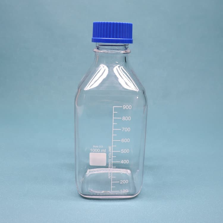
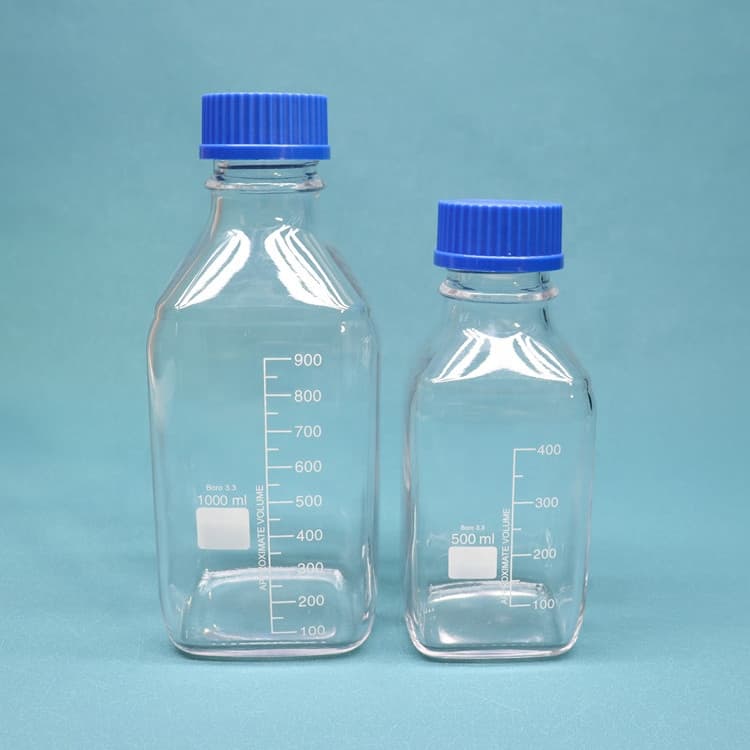
网页2018年3月1日 · Select the Nalgene dilution bottle that works for you. Nalgene dilution bottles make 1/10 and 1/100 dilutions simple. Graduated at 90mL and 99mL, with plenty
网页2019年5月24日 · Electron microscopy typically requires strong magnetic lenses in order to reach atomic resolution, prohibiting the possibility to measure magnetic materials. The
网页2023年1月4日 · Surface and bulk structures of cryogel disks were identified with scanning electron microscopy (SEM). The best MP release was found in 82.7% of the microsphere-cryogel composite system. Discover
网页The buffer is a vehicle for the fixative and requires certain properties: To maintain constant pH during the time of the fixation. The pH of the fixative in EM is adjusted using the buffer system chosen; e.g. range 6.5-8.0. The ionic constitution to prevent the extraction of cellular components, or the deposition of precipitates of fixative
网页Aqueous uranyl acetate has been extensively used as a superb staining reagent for transmission electron microscopy of biological materials. However, recent regulation of nuclear fuel material severely restricts its use even for purely scientific purposes. Since uranyl salts are hazardous due to biol
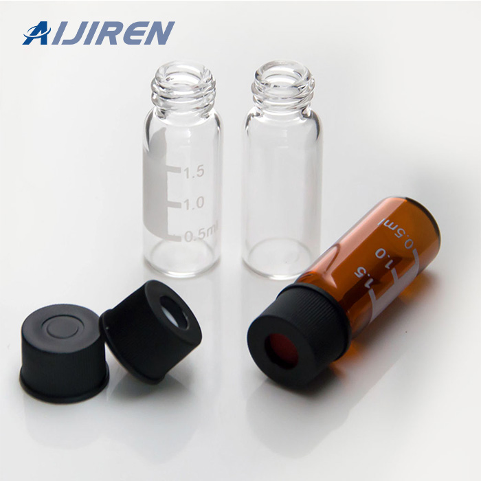
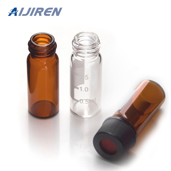
网页Shop Electron Microscopy Sciences Glass Sample Bottle With Plastic Snap Cap, 12 ml at Aijiren Techsci.com. Microscopy fixatives and stains – ANACC Methods and Materials
网页2015年7月16日 · 2. Negative staining technique. The classic processing of biological specimens observed in a TEM needs fixation, dehydration, sectioning and a selective “staining” of cell and tissue structures. “Staining”, a means of receiving coloured images, cannot be effectively used in conjunction with an electron microscope.
网页2013年3月7日 · 30. THE SCANNING ELECTRON MICROSCOPE • To directly visualise the surface topography of solid unsectioned specimens. • Probe scans the specimen in square raster pattern. • The first scanning electron microscope (SEM) debuted in 1938 ( Von Ardenne) with the first commercial instruments around 1965.
网页2019年5月24日 · Electron microscopy typically requires strong magnetic lenses in order to reach atomic resolution, prohibiting the possibility to measure magnetic materials. The authors here present a lens design
网页This third edition of Electron Microscopy: Methods and Protocols expands upon the previous editions with current, detailed protocols on biological and molecular research techniques based on TEM and SEM as well as other closely related imaging and analytical methods. With new chapters on conventional and microwave assisted specimen, cryo
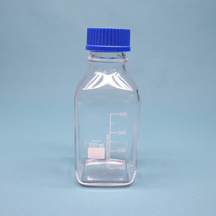
网页Square Bottles. Nalgene square bottles come in a variety of formats and materials to accommodate a wide range of application needs. Their square shape saves shelf and
网页2022年1月10日 · Immunofluorescence (IF) is a powerful method for visualizing intracellular processes, conditions and structures. IF preparations can be analyzed by various microscopy techniques (e.g. CLSM, Epifluorescence, TIRF, GSDIM), depending on the application or the researcher’s interest. Meanwhile, IF has become indispensable for a
网页2022年6月17日 · In this study, a new staining method for electron microscopic specimens is used. The method is based on the double staining of hematoxylin, which is widely used
网页EZ Pack™ Agarose Tablets. The quick dissolving (two minutes) EMS EZ Pack tablet contains 0.5g (500 mg) of EMS Agarose LE, eliminating the hands-on time and …